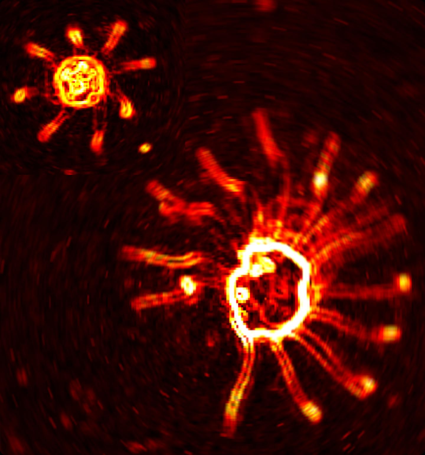
These are microscope images showing two species of algae which swim using tiny appendages known as flagella. Long before there were fish swimming in the oceans, tiny microorganisms were using long slender appendages called cilia and flagella to navigate their watery habitats. Now, new research reveals that species of single-celled algae coordinate their flagella to achieve a remarkable diversity of swimming gaits.
When it comes to four-legged animals such as cats, horses and deer, or even humans, the concept of a gait is familiar, but what about unicellular green algae with multiple limb-like flagella? The latest discovery, published in the journal Proceedings of the National Academy of Sciences, shows that despite their simplicity, microalgae can coordinate their flagella into leaping, trotting or galloping gaits just as well.
Many gaits are periodic: whether it is the stylish walk of a cat, the graceful gallop of a horse, or the playful leap of a springbok, the key is the order or sequence in which these limbs are activated. When springboks arch their backs and leap, or 'pronk', they do so by lifting all four legs simultaneously high into the air, yet when horses trot it is the diagonally opposite legs that move together in time.
In vertebrates, gaits are controlled by central pattern generators, which can be thought of as networks of neural oscillators that coordinate output. Depending on the interaction between these oscillators, specific rhythms are produced, which, mathematically speaking, exhibit certain spatiotemporal symmetries. In other words, the gait doesn't change when one leg is swapped with another - perhaps at a different point in time, say a quarter-cycle or half-cycle later.
It turns out the same symmetries also characterise the swimming gaits of microalgae, which are far too simple to have neurons. For instance, microalgae with four flagella in various possible configurations can trot, pronk or gallop, depending on the species.
"When I peered through the microscope and saw that the alga was performing two sets of perfectly synchronous breaststrokes, one directly after the other, I was amazed," said the paper's first author Dr Kirsty Wan of the Department of Applied Mathematics and Theoretical Physics (DAMTP) at the University of Cambridge. "I realised immediately that this behaviour could only be due to something inside the cell rather than passive hydrodynamics. Then of course to prove this I had to expand my species collection."
The researchers determined that it is in fact the networks of elastic fibres which connect the flagella deep within the cell that coordinate these diverse gaits. In the simplest case of Chlamydomonas, which swims a breaststroke with two flagella, absence of a particular fibre between the flagella leads to uncoordinated beating. Furthermore, deliberately preventing the beating of one flagellum in an alga with four flagella has zero effect on the sequence of beating in the remainder.
However, this does not mean that hydrodynamics play no role. In recent work from the same group, it was shown that nearby flagella can be synchronised solely by their mutual interaction through the fluid. There is a distinction between unicellular organisms for which good coordination of a few flagella is essential, and multicellular species or tissues that possess a range of cilia and flagella. In the latter case, hydrodynamic interactions are much more important.
"As physicists our instinct is to seek out generalisations and universal principles, but the world of biology often presents us with many fascinating counterexamples," said Professor Ray Goldstein, Schlumberger Professor of Complex Physical Systems at DAMTP, and senior author of the paper. "Until now there have been many competing theories regarding flagellar synchronisation, but I think we are finally making sense of how these different organisms make best use of what they have."
The findings also raise intriguing questions about the evolution of the control of peripheral appendages, which must have arisen in the first instance in these primitive microorganisms.
Source: University of Cambridge
 Print Article
 Mail to a Friend
