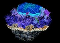
This 3-D reconstruction of a human skin cell was produced by electron tomography and shows organelles in different colours: regions of cell-cell contact (sandy brown), nucleus and nuclear envelope (blue) with pores (red), microtubules (green), mitochondria (purple), endoplasmic reticulum (steel blue). Credit: Achilleas Frangakis EMBL Seeing proteins in their natural environment and interactions inside cells has been a long-standing goal. Using an advanced microscopy technique called cryo-electron tomography, researchers from the European Molecular Biology Laboratory [EMBL] have visualised proteins responsible for cell-cell contacts for the first time. In this week’s issue of Nature they publish the first 3D image of human skin at molecular resolution and reveal the molecular Velcro-like organisation that interlinks cells.
“This is a real breakthrough in two respects,” says Achilleas Frangakis, group leader at EMBL. “Never before has it been possible to look in three dimensions at a tissue so close to its native state at such a high resolution. We can now see details at the scale of a few millionths of a millimetre. In this way we have gained a new view on the interactions of molecules that underlie cell adhesion in tissues – a mechanism that has been disputed over decades.” __IMAGE_2
So far, the only information available about a protein’s position and interactions in a cell was based on either light microscopy images at poor resolution or techniques that remove proteins from their natural context. Frangakis and his group have been developing a technique called cryo-electron tomography, with which a cell or tissue is instantly frozen in its natural state and then examined with an electron micro-scope. Electron microscopy normally requires tissue to be treated with chemicals or coated in metal, a procedure that disturbs the natural state of a sample. With cyro-electron tomography, images are taken of the untreated sample from different directions and assembled into an accurate 3D image by a computer.
The researchers applied this technique to observe proteins that are crucial for the integrity of tissues and organs like the skin and the heart, but also play an important role in cell proliferation. These proteins, called cadherins, are anchored in cell membranes and interact with each other to bring cells close together and interlink them tightly.
“We could see the interaction between two cadherins directly, and this revealed where the strength of human skin comes from,” says Ashraf Al-Amoudi, who carried out the work in Frangakis’ lab. “The trick is that each cadherin binds twice: once to a molecule from the juxtaposed cell, and once to its next-door neighbour. The system works a bit like specialised Velcro and establishes very tight contacts between cells.”
The new insights into the cadherin system broadens the understanding of structural aspects of cell adhesion and shed light on other crucial processes such as cell proliferation. The technical advances achieved in cryo-electron tomography of frozen sections open up new possibilities to study more systems at native conditions with molecular resolution.
Source : European Molecular Biology Laboratory
 Print Article
 Mail to a Friend
