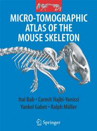
Cover of the new book by Professor Itai Bab and associates at the Hebrew University of Jerusalem on the skeletal structure of the mouse. A new book by researchers at the Hebrew University of Jerusalem that details the skeletal structure of the mouse demonstrates a surprising similarity between mice and humans.
The book,”Micro-Tomorgrphic Atlas of the Mouse Skeleton,” was authored by a team from the Hebrew University Bone Laboratory consisting of Prof. Itai Bab, head of the laboratory; Dr. Carmit Hajbi-Yonissi and Dr. Yankel Gabet. Also participating in the writing of the book was Dr. Ralph Müller of the ETH of Zürich. The book, published by Springer of New York, provides great visual detail of the mouse skeletal structure, utilizing the technology of micro-tomographic imaging.
The authors observe that there are many areas of comparison between the mouse and human skeleton, with the exception of the facial, hand and foot bones. This is very important because, for example, mice, like people, suffer from osteoporosis. The significance of this lies in that research on osteoporosis in mice can have great relevance for and applications to humans. The same is true in relation to other problems related to fractures, skeletal development and illnesses, including testing of drugs.
According to Prof. Bab, earlier anatomical works on the mouse skeleton were based on visual observations only and are insufficient for purposes of modern research. Those books contained inexact material, insufficient detail, and only partial descriptions of the various skeletal components, said Bab.
Prof. Bab explained that in the last decade the new computerized micro-tomographic technology which has been developed provides excellent skeletal imaging. Using this technology, one can see two and three-dimensional images which show details down to the six-thousandth of a millimeter. The new atlas presents almost 200 such two and three-dimensional images, showing all external portions of the mouse skeleton and also the internal anatomy of the bones.
Also presented are three-dimensional images of the relationships between the bones at various joint positions. One chapter of the book, based on measurements of skeletons at various ages, describes the development of the skeleton and its aging and also the differences between males and females.
The micro-tomographic equipment used by the researchers was purchased by the Hebrew University. The purchase was partially supported by a grant from the Israel Science Foundation.
Source : The Hebrew University of Jerusalem
 Print Article
 Mail to a Friend
