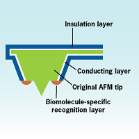
By adding electrodes to the tip of an atomic force microscope, researchers created a tool that can monitor many activities at the same time."Usually people image topography and then measure the biological activity," said Christine Kranz, research scientist at Georgia Tech "But if you think about having biological material, it's changing with time. So scanning for these sequentially may mean that the structure and activity level of your sample has changed and you're not looking at the same condition of your sample anymore."
Using an AFM as the base, researchers added micro and nano-electrodes into the tip. This allows researchers to get biological and chemical information via scanning electrochemical microscopy (SECM) simultaneously with topographical information provided by AFM.
"The problem with conventional AFM imaging is that you get topographical information, but only limited information on the chemical processes occurring at the cell surface," said Kranz. "With SECM, you can get information on electro-activity, but this information may be convoluted with the topographical information. Also SECM still suffers from limited resolution. We combined the two techniques to give us high resolution topography as well as the chemistry that's going on at the cell surface."
Researchers tested their new technique by integrating biosensors for glucose into AFM tips. They imaged, as a synthetic model, glucose transport through track-etched membranes. In another test, a biosensor based on horse radish peroxidase was integrated into the AFM tip. They were able to faithfully measure the chemical activity and image the process to a resolution of 200 nanometers.
The tool promises to be valuable for a wide range of biomedical and biotechnological applications. In an NIH-funded project in collaboration with Emory University, Kranz and Boris Mizaikoff are currently using this technique to study cystic fibrosis and the role errors in regulating adenosine triphosphate (ATP), a chemical involved in transporting energy to cells, might play in the disease.
"The system's modular design allows it to be adapted for many uses," said Kranz. Researchers at the Applied Sensors Laboratory at Georgia Tech are testing integrating optical microscopy with the AFM. Another project adds an infrared sensor, while yet another adds a pH sensor. These sensors could also be combined to have three or more sensors on a single instrument because the technology is in the modified AFM tip.
"The technology is very flexible and can be adapted to sense for a wide range of biological systems and processes. Using this technique, we can get a more complete picture of what is going on in a given biological system," said Kranz.
The research team consisted of Kranz, Mizaikoff, and former postdoc Angelika Kueng from Georgia Tech. Focused ion beam milling was done by Alois Lugstein and Emmerich Bertagnolli from the Vienna University of Technology. Since January 2005, Georgia Tech has established a Focused Ion Beam Center led by Mizaikoff enabling tip fabrication, characterization, and measurements on campus.
Source : Georgia Institute of Technology
 Print Article
 Mail to a Friend
